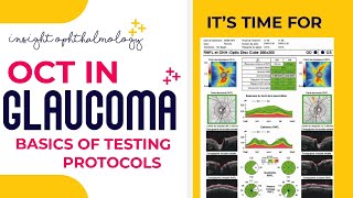
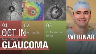
Lecture: How to Interpret OCT Findings in the Diagnostic Evaluation of Glaucoma
Video Not Working? Fix It Now
Optical coherence tomography (OCT) has become an indispensable tool in the diagnostic evaluation of glaucoma. However, deciding what regions (e.g., optic disc region vs. macula) and what parameters (e.g., retinal nerve fiber layer (RNFL) thickness vs. ganglion cell inner plexiform layer (GCIPL) thickness vs. neuroretinal rim width) to measure can often be confusing. False positives and false negatives in OCT reports are common. This webinar will walk through examples from different OCT models and elaborate correct interpretation of OCT findings to aid diagnosis and monitoring of glaucoma.
Lecturer: Dr. Chris Leung, Head of Dept., Clinical Professor, Dept. of Ophthalmology, The University of Hong Kong
Comment